(click to enlarge)
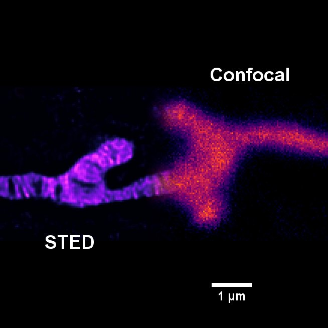
H9c2 cell labeled with Live ORANGE Mito dye. The same field was captured with conventional confocal imaging and STED super resolution microscopy and then images are blended for comparison. Note that the mitochondrial inner membrane network (cristae) is resolvable with STED but not confocal. Sample courtesy of Nathan Alder and Subrata Biswas.