(click to enlarge)
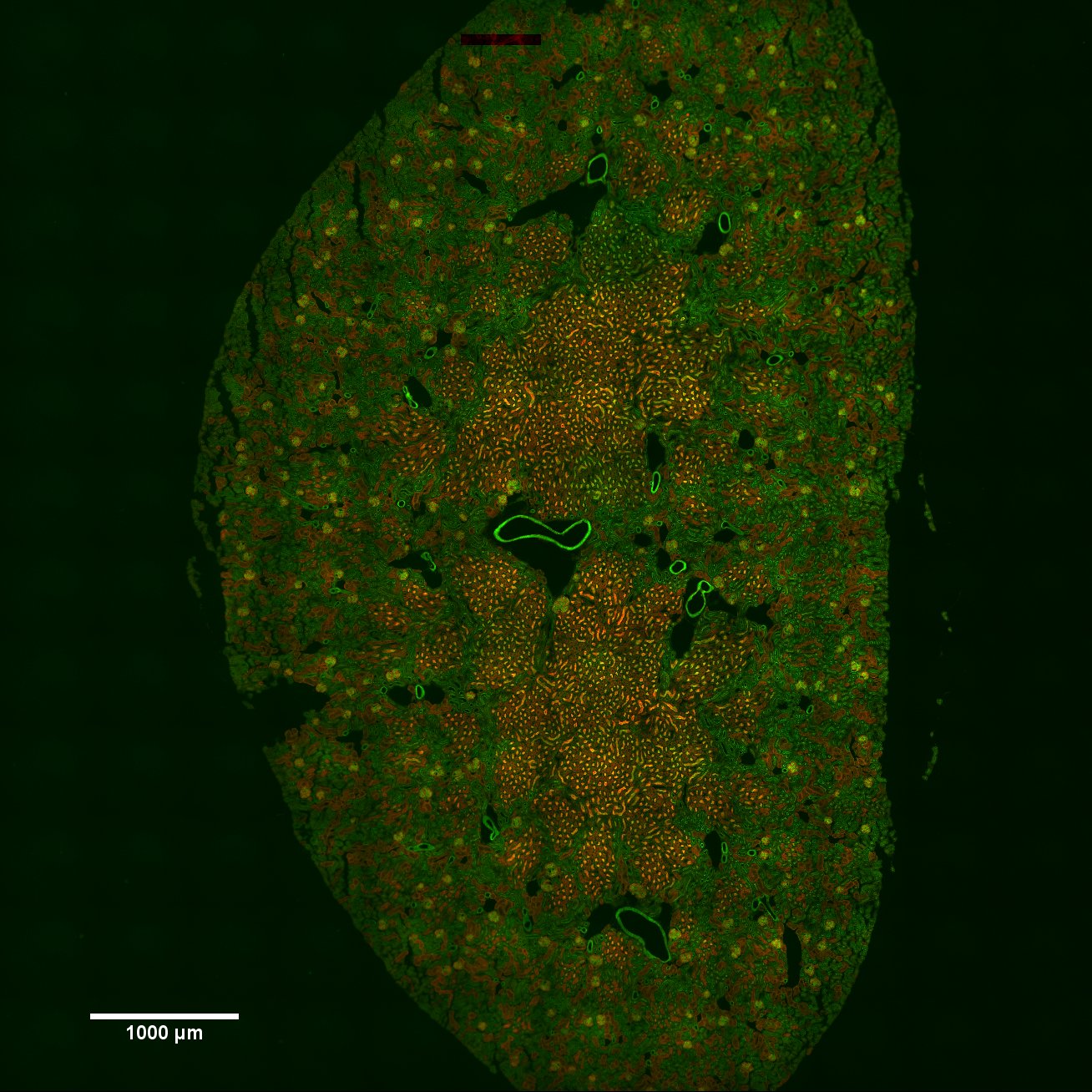
Stitched image of an entire mouse kidney section captured with widefield fluorescence microscopy of WGA (green) and phalloidin (red). The image is assembled from a 12 X 12 grid of images captured with a 20X objective lens on the Andor System. Courtesy of Chris O’Connell.