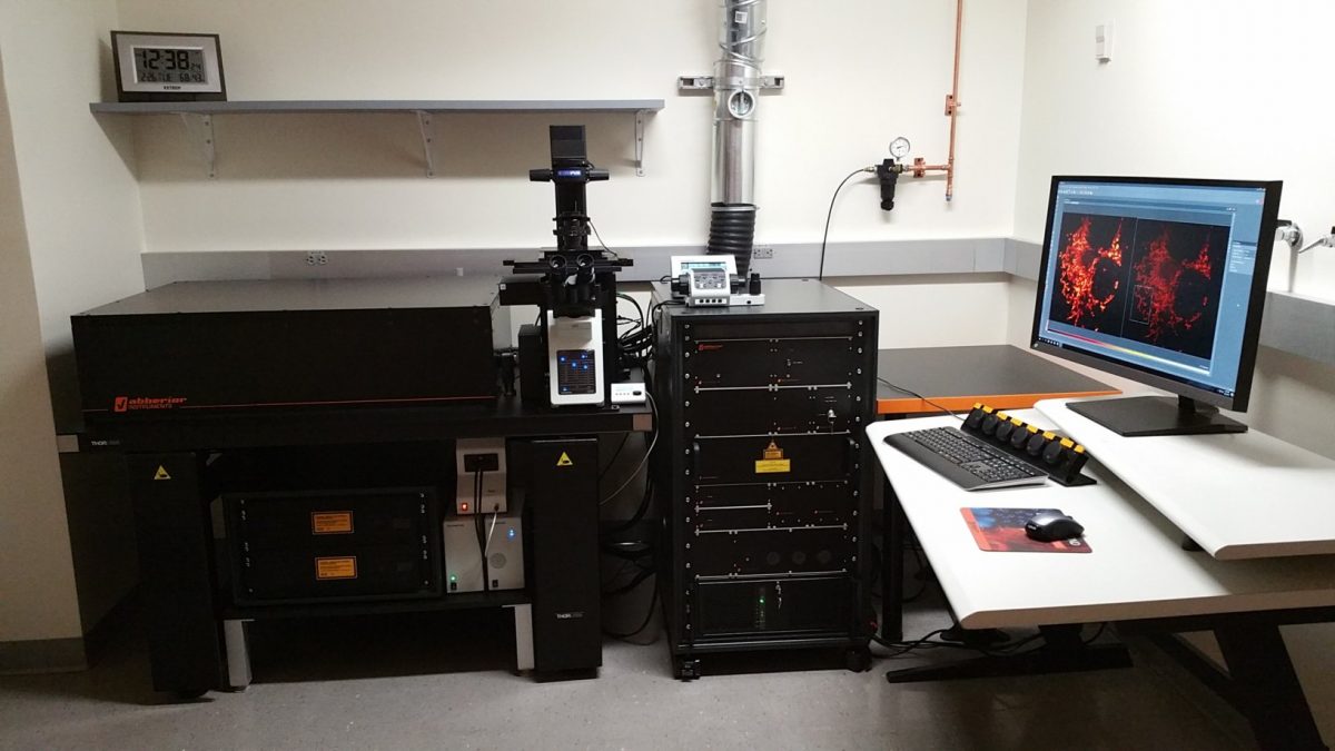
Overview: This is an inverted confocal microscope that uses stimulated emission depletion (STED) of fluorescent dyes to obtain super resolved images. There are four pulsed excitation lasers and two pulsed STED depletion lasers for imaging. The instrument is capable of 2D or 3D super resolution imaging using spatial light modulators to shape the depletion beams in the x-y and z axes. The instrument is housed within the Bioscience Electron Microscopy Laboratory located in the Biology and Physics Building (BPB), room G11. Click here for rate information.
Assisted use with the Facility Director is preferred. At the discretion of the Director, users can be trained to operate the system independently if they have demonstrated mastery of conventional confocal microscopy (Leica SP8, Nikon A1R, or equivalent) and have a substantial need for STED imaging in their project.
Samples for STED imaging should be prepared with labels, coverslips, and mounting medium optimized for the technique. Consult our sample prep guide for recommendations.
This microscope was acquired with a NIH shared instrumentation grant. Manuscripts including data from the Abberior STED system must include an acknowledgement of NIH grant S10OD023618 awarded to Christopher O’Connell.
Microscope:
- Inverted Stand Olympus IX83
- Motorized x-y stage
- Physik Instrumente piezo z stage insert
Lasers:
Excitation
- 405 nm CW (non-STED imaging)
- 440 nm pulsed
- 485 nm pulsed
- 561 nm pulsed
- 640 nm pulsed
STED Depletion
- 595 nm pulsed
- 775 nm pulsed
Scanner:
- Continuously adjustable QUADScan galvo scanner (up to 2600 Hz line scanning)
Objective Lenses:
- 10X/0.3 UPLFLN10X2
- 20x/0.75 UPLSAPO20
- 40x/0.1.3 UPLFNL40 oil immersion
- 60x/1.2 UPLSAPO60 water immersion
- 100x/1.40 UPLANSAPO oil immersion
Detectors:
- Four filter-based APD detectors
Software:
- Abberior Impsector for acquisition and visualization
